Wavefront Engineering Microscopy
The research focus of the group has a dual technological and neuroscientific objective. Firstly, it involves advancing all-optical methodologies by integrating single and multiphoton excitation with spatiotemporal wave-front shaping, holographic illumination, compressed sensing, and probe engineering. Secondly, utilizing this unique combination to investigate, at unprecedented precision, neuronal circuits crucial for visual perception.
Presentation
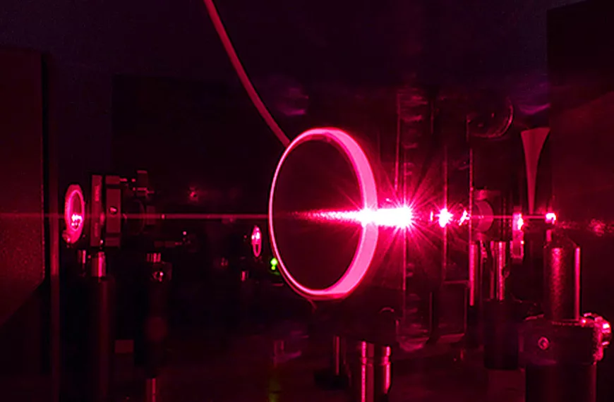
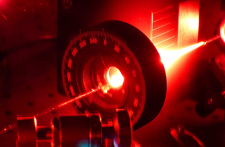
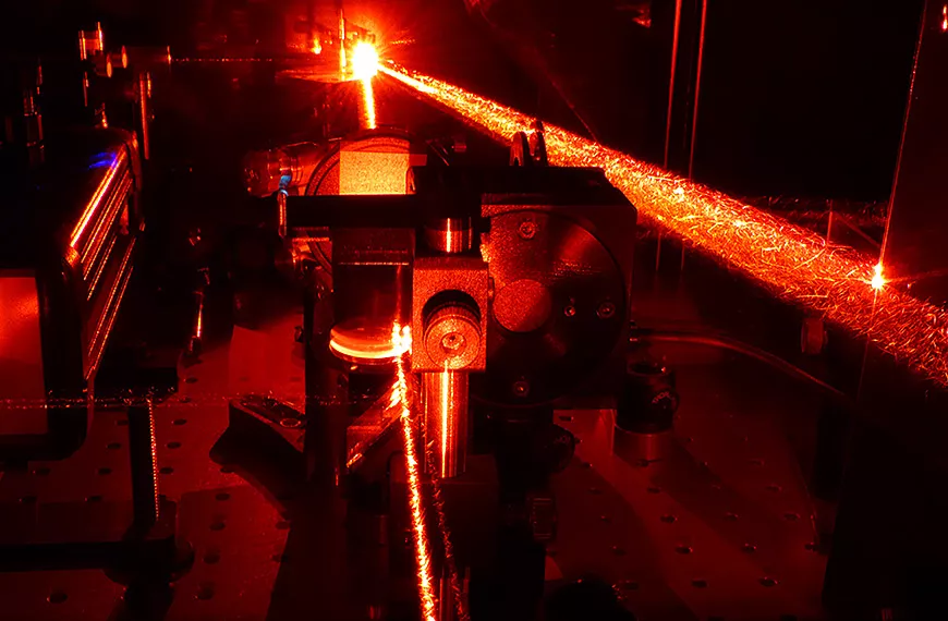
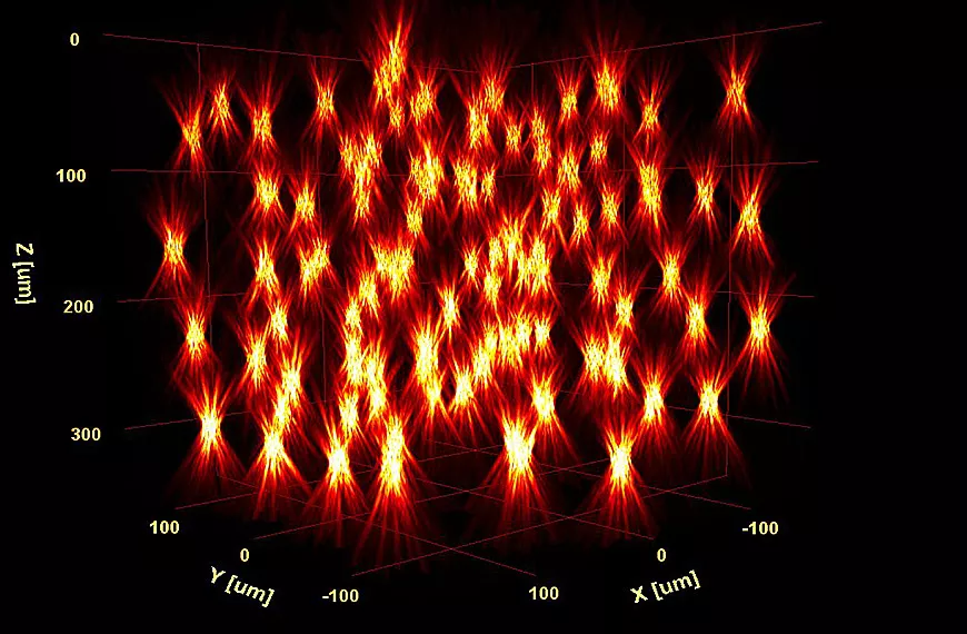
The Wave-front engineering Microscopy group is an interdisciplinary team composed by physicists, engineers, biophysicists and neurophysiologists. The group has pioneered the use of wave front shaping for all-optical brain manipulation, in a few very early and breakthrough papers, demonstrating spatiotemporal wave front shaping approaches such as computer-generated holography, generalized phase contrast and temporal focusing, and showing that such methods can be used to sculpt the excitation volume with a shape perfectly tailored on the selected target.
The combination of these approaches with newly developed optogenetics actuators and high-power amplified lasers enables the control of single or multiple targets independently in space and time in mm3 volume at cellular resolution and millisecond temporal precision. We termed this combination of approaches, circuits optogenetics The unprecedented spatiotemporal precision of circuit optogenetics is currently being utilized by my team and other labs worldwide in a wide range of experimental paradigms. Among the collaborative projects of my group with other neuroscience laboratories, we can mention the use of circuits optogenetics for high-throughput connectivity mapping of neuronal circuits in zebrafish or the visual cortex, for the optical manipulation of retinal circuits, and for the in vivo demonstration of hub cells in neuronal circuits during development.
Combining single photon holography and endoscopy we were also able to demonstrate simultaneous photostimulation and functional imaging with near-cellular resolution in freely moving mice and more recently we have been able to make this approach also compatible with two-photon excitation.
We have recently demonstrated that scanless two-photon microscopy also provides important advantages for two-photon voltage imaging. Specifically, we have shown that, in combination with temporal focusing, parallel illumination enables high-contrast, high-resolution in vitro and in vivo voltage imaging in densely labelled preparation (mice cortex and zebrafish larvae) expressing GFP and rhodopsin based genetically encoded voltage indicator.
Our current research activity focuses on advancing even further the development of these technologies and on the use of the developed technologies for the investigation of the mechanisms regulating functional connectivity and signal processing across the main visual pathways in mice, non-human primates and zebrafish larvae.

Research areas
- Multiphoton scanless optogenetics
- Two-photon holographic endoscopy
- Two-photon scanless voltage imaging
- All-optical circuits manipulation: circuits optogenetics
- Holographic vision restauration
- Dissemination
Team members
Scientific publications
Below you will find the latest scientific publications in this field: Wavefront Engineering Microscopy.


